Digital X-ray in Kota
Digital X-ray
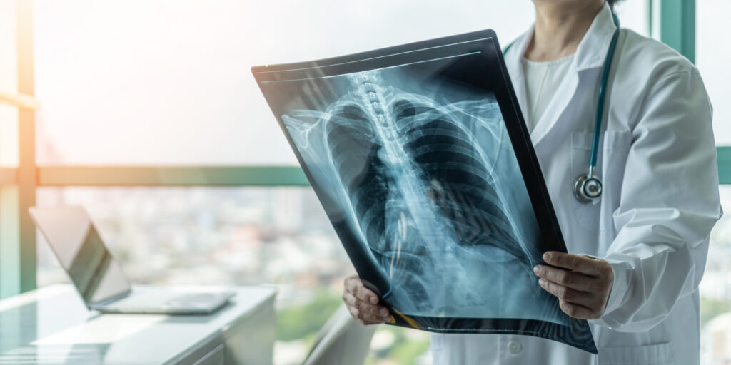
X-rays are a form of electromagnetic radiation, just like visible light. In a health care setting, a machines sends are individual x-ray particles, called photons. These particles pass through the body. A computer or special film is used to record the images that are created.
Structures that are dense (such as bone) will block most of the x-ray particles, and will appear white. Metal and contrast media (special dye used to highlight areas of the body) will also appear white. Structures containing air will be black, and muscle, fat, and fluid will appear as shades of gray.
How the Test is Performed
The test is performed in a hospital radiology department by an x-ray technologist. The positioning of the patient, x-ray machine, and film depends on the type of study and area of interest. Multiple individual views may be requested.
Much like conventional photography, motion causes blurry images on radiographs, and thus, patients may be asked to hold their breath or not move during the brief exposure (about 1 second).
How to Prepare for the Test
Inform the health care provider prior to the exam if you are pregnant, may be pregnant, If abdominal studies are planned and you have had a barium contrast study (such as a barium enema, upper GI series, or barium swallow) in the last 4 days, the test may be delayed until the contrast has fully passed.
You will remove all jewelry and wear a hospital gown during the x-ray examination because metal and certain clothing can obscure the images and require repeat studies.
Contact Us to get more Information About Our Laboratory
How the Test Will Feel
There is no discomfort from x-ray exposure. Patients may be asked to stay still in awkward positions for a short period of time.
- Risks
For most conventional x-rays, the risk of cancer or defects due to damaged ovarian cells or sperm cells is very low. Most experts feel that this low risk is largely outweighed by the benefits of information gained from appropriate imaging. X-rays are monitored and regulated to provide the minimum amount of radiation exposure needed to produce the image.
Young children and fetuses are more sensitive to the risks of x-rays. Women should tell health care providers if they think they are pregnant.
- Alternative Names
Radiography
Benefits of Digital X-ray
Digital X-rays offer several advantages over traditional X-rays. These include:
- Faster Results: Images are available almost immediately, allowing quicker diagnosis and treatment.
- Improved Image Quality: Digital images can be enhanced, zoomed, or adjusted for better clarity.
- Reduced Radiation Exposure: Digital X-ray technology typically requires lower doses of radiation compared to traditional film X-rays.
- Easy Storage and Sharing: Digital files can be easily stored and shared with other healthcare providers if needed.
- Environmentally Friendly: No need for chemicals used in film development, making the process more eco-friendly.
Applications of Digital X-ray
Digital X-rays are commonly used for:
- Chest X-rays: To diagnose conditions such as pneumonia, heart problems, or lung diseases.
- Bone Imaging: To detect fractures, infections, or bone abnormalities.
- Dental X-rays: Used to examine teeth, gums, and surrounding tissues.
- Mammography: A special type of X-ray for breast cancer screening.
- Fluoroscopy: A technique that uses X-rays to obtain real-time moving images of the interior of the body, such as during a barium swallow test.
Why Choose Our Lab for Digital X-ray Services ?
At Dr. Vaya’s Lab in Kota, we use state-of-the-art digital X-ray technology to ensure accurate results and high-quality care. Our experienced technicians work closely with radiologists to provide comprehensive imaging services. Here’s why patients trust us:
- Advanced Equipment: Our lab is equipped with modern X-ray machines that provide precise imaging while minimizing radiation exposure.
- Experienced Team: Our certified technologists and radiologists have years of experience in diagnostic imaging.
- Patient-Centered Care: We prioritize patient comfort, ensuring a smooth and efficient X-ray experience.
- Convenient Appointments: We offer flexible scheduling to accommodate your needs.
If you're looking for reliable digital X-ray services in Kota, Dr. Vaya’s Lab is here to help. For more information or to schedule an appointment, please contact us today.
Frequently Asked Questions
A digital X-ray is an advanced imaging technique that uses digital sensors to capture images of the inside of the body. It replaces traditional film X-rays and provides faster, clearer, and more detailed results.
Digital X-rays use electronic sensors instead of film, produce images instantly, require less radiation, and allow for easier storage and sharing of images.
They are used to diagnose and monitor conditions such as fractures, infections, tumors, lung issues, dental problems, and more.
Yes, digital X-rays are safe. They use up to 80% less radiation than traditional X-rays, making them safer for patients, including children and pregnant women (with proper precautions).
- The process is quick, usually taking 10-15 minutes per body part. Results are often available immediately or within 24-48 hours.
X-ray Categories
| ABDOMEN STANDING |
| ABDOMEN AP/LAT |
| APIOGRAM |
| KNEE STANDING BOTH KNEE AP |
| BARIUM ANIMA |
| BARIUM MEAL FLOW THROUGH ( BMFT) |
| C. SPINE AP/ LAT |
| CHEST WITH ABD. |
| CHEST PA |
| CT. SCAN REPORTING |
| CYSTOGRAM |
| CHEAT AP |
| D.L. SPINE AP/LAT |
| PELIVIS AP |
| ELBOW AP/ LAT |
| BOTH KNEE AP/ LAT |
| CERVICAL DORSAL SPINE AP/LAT |
| FPA STANDING |
| IVP WITH CYSTROGRAM |
| HAND FOREARM ELBOW AP/LAT |
| LT. WRIST AP / LAT |
| NECK LAT VIEW |
| NOSE AP/ LAT |
| THIGH AP /LAT |
| KNEE AP LAT |
| KNEE B/L AP / LAT ( BOTH) |
| ANKLE AP/ LAT |
| LEG AP/ LAT. |
| BARIUM SWALLOW |
| ANKLE LAT ( BOTH ANKLE) |
| FEMUR AP/ LAT ( BOTH) |
| FOOT AP/ LAT ( BOTH) |
| HAND AP ( BOTH) |
| HAND LAT. ( BOTH) |
| HAND AP/ LAT ( BOTH HAND) |
| HEEL AP/ LAT. ( BOTH) |
| BOTH HIP AP |
| HIP AP/ LAT. |
| KNEE AP ( BOTH) |
| BOTH LEG AP/LAT |
| BOTH MASTOIDS |
| BOTH WRIST AP |
| C.SPINE LAT |
| CHEST LAT. |
| CYSTOURETROGRAM |
| D.L. SPINE AP/LAT/ OBLIQ |
| L.S. SPINE WITH L.S. JUNCTION |
| T.M JOINT OPEN CLOSE MOUTH |
| CLAVICAL AP |
| HUMEROUS AP/ LAT |
| KNEE AXIAL |
| SHOULDER AP/ AXIAL |
| WRIST AP/ LAT/ OBLIQ |
| MANDIBLE AP/ LAT |
| MANDIBLE PA |
| NOSAL BONE |
| PELIVIS AP /LAT/ FROG |
| FOOT AP/ LAT |
| HAND AP /LAT |
| HEEL AP/ AXIAL |
| HEEL LAT/ AXIAL |
| SHOULDER AP / LAT |
| WRIST AP/ LAT |
| S.I JOINT |
| SACRO COCCYX AP/LAT |
| SKULL AP |
| SKULL AP/LAT |
| SKULL LAT |
| SOFT TISSUE NECK |
| PELVIC BOTH HIP AP |
| ORBIT AP/ LAT |
| PELVIS Lat |
| WRIST AP |
| FEMUR WITH KNEE AP/ LAT |
| SACRUM AP/ LAT. |
| SACRUM AP |
| SACRUM LAT |
| LOOPOGRAM |
| FESTULOGRAM |
| GASTROGRAFFIN SWALLOW |
| GENITOGRAM |
| HAND INCLUDING WRIST JT. AP |
| HEEL B/L MEDIAL VIEW |
| HSG |
| IVP |
| KNEE WITH UPPER LEG AP/ LAT |
| KUB |
| LAT. CEPHALOGRAM |
| MCU |
| MESTOID |
| OPG |
| P.N.S ( WATER VIEW) |
| PCN GRAM |
| PELVIS WITH BOTH HIP JOINT |
| RGU |
| RGU/ MCU |
| THUMB AP/ LAT |
| THUMB AP |
| THUMB LAT |
| SACRUM LAT |
| SINOGRAM |
| TMJ VIEW |
| VCUG |