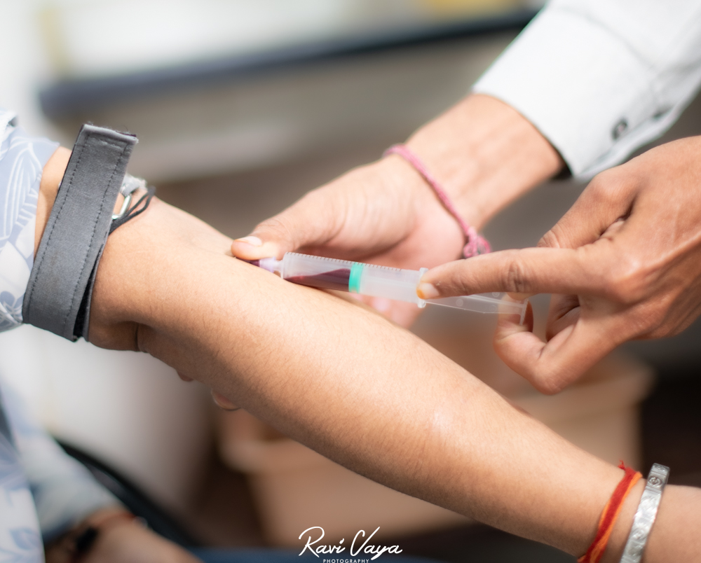
In the ever-evolving field of medical diagnostics, minimally invasive techniques have revolutionized the way we approach patient care. One such area where these advancements have made significant strides is in urology. Fluoroscopic imaging, in particular, has become an indispensable tool for urologists, allowing for real-time visualization of the urinary tract with minimal discomfort to the patient. This article delves into the world of fluoroscopic imaging in urology, exploring its applications, benefits, and the role of diagnostic centers in Kota in providing these cutting-edge services.
Understanding Fluoroscopic Imaging
Fluoroscopy is a type of medical imaging that uses X-rays to obtain real-time moving images of the interior of an object. In urology, this technique is particularly valuable for visualizing the urinary tract, including the kidneys, ureters, bladder, and urethra. The ability to see these structures in motion provides urologists with crucial information that static imaging methods might miss.
How Fluoroscopy Works in Urological Diagnostics
- Continuous X-ray Beam: A fluoroscope machine emits a continuous X-ray beam that passes through the patient’s body.
- Image Intensifier: The X-rays that pass through the body are captured by an image intensifier, which converts them into a visible light image.
- Video Camera: This visible light image is then captured by a video camera and displayed on a monitor in real-time.
- Contrast Agents: Often, contrast agents are used to enhance the visibility of specific structures within the urinary system.
- Dynamic Visualization: The real-time nature of fluoroscopy allows urologists to observe the function and flow within the urinary tract.
Applications of Fluoroscopic Imaging in Urology
Fluoroscopy has a wide range of applications in urological diagnostics and procedures. Some of the key areas where this technology proves invaluable include:
- Voiding Cystourethrogram (VCUG): This procedure uses fluoroscopy to examine the bladder and urethra during urination, helping diagnose conditions like vesicoureteral reflux.
- Retrograde Pyelogram: Fluoroscopy guides the insertion of a catheter into the ureter to inject contrast dye, allowing visualization of the upper urinary tract.
- Percutaneous Nephrolithotomy (PCNL): Fluoroscopic guidance is crucial in this minimally invasive procedure for removing kidney stones.
- Ureteral Stent Placement: Real-time imaging helps ensure accurate placement of ureteral stents to relieve obstructions.
- Urodynamic Studies: Fluoroscopy can be used in conjunction with urodynamic testing to assess bladder and urethral function.
Benefits of Fluoroscopic Imaging in Urology
The adoption of fluoroscopic imaging techniques in urology offers numerous advantages:
- Minimally Invasive: Fluoroscopy allows for diagnostic procedures with minimal discomfort to the patient.
- Real-Time Visualization: The ability to see structures in motion provides valuable functional information.
- Reduced Radiation Exposure: Compared to traditional X-rays, fluoroscopy can often be performed with lower overall radiation doses.
- Improved Accuracy: Real-time imaging enhances the precision of diagnoses and interventions.
- Versatility: Fluoroscopy can be used for both diagnostic imaging and to guide therapeutic procedures.
The Role of Diagnostic Centers in Kota
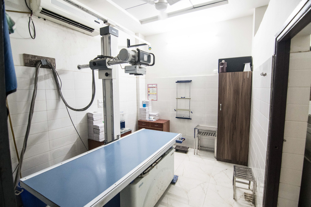
As fluoroscopic imaging becomes increasingly important in urological care, the role of specialized diagnostic centers becomes crucial. In Kota, a city known for its medical facilities, several diagnostic points in Kota offer advanced fluoroscopic services. These centers play a vital role in:
- Providing Access to Advanced Technology: Modern diagnostic centers in Kota invest in state-of-the-art fluoroscopic equipment, ensuring patients have access to the latest diagnostic tools.
- Specialized Expertise: Technicians and radiologists at these centers are trained specifically in fluoroscopic techniques, ensuring high-quality imaging and interpretation.
- Collaborative Care: Diagnostic centers in Kota often work closely with urologists, providing a seamless experience for patients from imaging to treatment.
- Patient Education: These centers play a crucial role in educating patients about the procedures, addressing concerns, and ensuring comfort during the imaging process.
- Research and Innovation: Many diagnostic points in Kota participate in research, contributing to the advancement of fluoroscopic techniques in urology.
Choosing the Right Diagnostic Centre in Kota
For patients and healthcare providers looking to utilize fluoroscopic imaging services, selecting the right diagnostic center is crucial. Here are some factors to consider:
- Technology and Equipment: Look for centers with modern, well-maintained fluoroscopic equipment.
- Expertise of Staff: Ensure the center has radiologists and technicians experienced in urological fluoroscopy.
- Accreditation and Certifications: Choose centers that meet national standards for imaging quality and safety.
- Patient Comfort: Consider facilities that prioritize patient comfort and offer clear explanations of procedures.
- Collaborative Approach: Opt for centers that work closely with referring urologists for comprehensive care.
Advancements in Fluoroscopic Imaging
The field of fluoroscopic imaging in urology continues to evolve, with several exciting developments on the horizon:
- Digital Subtraction Angiography (DSA): This technique enhances the visibility of blood vessels, useful in diagnosing vascular abnormalities in the urinary system.
- 3D Fluoroscopy: Advanced systems can create three-dimensional images, providing more detailed information for complex cases.
- Dose Reduction Technologies: Newer fluoroscopic systems incorporate features to minimize radiation exposure while maintaining image quality.
- AI-Assisted Interpretation: Artificial intelligence algorithms are being developed to assist in the interpretation of fluoroscopic images, potentially improving diagnostic accuracy.
- Fusion Imaging: Combining fluoroscopy with other imaging modalities like ultrasound or CT for more comprehensive diagnostics.
Preparing for a Fluoroscopic Procedure
For patients scheduled for a fluoroscopic examination at a Kota diagnostic center, proper preparation is key to ensuring a successful procedure:
- Follow Instructions: Adhere to any specific instructions provided by the diagnostic center regarding food, drink, or medication restrictions.
- Inform About Medications: Notify the center about any medications you’re taking, especially those that might affect urinary function.
- Discuss Allergies: Inform the staff about any allergies, particularly to contrast agents or iodine.
- Wear Comfortable Clothing: Choose loose, comfortable clothing that’s easy to change out of for the procedure.
- Stay Hydrated: Unless instructed otherwise, maintaining good hydration can be helpful for certain urological fluoroscopic procedures.
The Future of Fluoroscopic Imaging in Urology
As technology continues to advance, the future of fluoroscopic imaging in urology looks promising:
- Enhanced Image Quality: Ongoing improvements in detector technology promise even clearer, more detailed images.
- Reduced Radiation Exposure: Future systems may offer equivalent or superior imaging with even lower radiation doses.
- Integration with Other Technologies: Expect to see more seamless integration of fluoroscopy with other diagnostic and treatment modalities.
- Portable Systems: Advancements may lead to more portable fluoroscopic systems, increasing accessibility in various healthcare settings.
- Personalized Imaging Protocols: AI-driven systems might tailor imaging parameters to individual patient characteristics for optimal results.
Addressing Common Concerns
While fluoroscopic imaging is generally safe and well-tolerated, patients often have concerns. Here are some common questions addressed:
- Radiation Exposure: Modern fluoroscopic systems use the lowest possible radiation dose to obtain diagnostic images. The benefits of the procedure typically outweigh the minimal risks associated with radiation exposure.
- Contrast Agent Reactions: While rare, some patients may experience allergic reactions to contrast agents. Diagnostic centers in Kota are well-equipped to handle such situations.
- Procedure Duration: Most fluoroscopic procedures in urology are relatively quick, typically lasting 15 to 30 minutes.
- Discomfort: While some procedures may cause mild discomfort, they are generally well-tolerated. The staff at Kota diagnostic centers prioritize patient comfort throughout the process.
- Results Availability: In many cases, preliminary results are available immediately after the procedure, with detailed reports following shortly after.
Conclusion
Fluoroscopic imaging has become an indispensable tool in modern urology, offering minimally invasive diagnostic and treatment options that benefit both patients and healthcare providers. As diagnostic points in Kota continue to invest in advanced fluoroscopic technology and expertise, patients in the region have access to world-class urological care.
The combination of real-time visualization, reduced invasiveness, and improved diagnostic accuracy makes fluoroscopy a cornerstone of urological diagnostics. As technology continues to advance, we can expect even more exciting developments in this field, further enhancing patient care and outcomes.
FAQs about Fluoroscopic Imaging in Urology
1. Does Dr. Vaya’s Lab, Kota offer fluoroscopic imaging services for urological diagnostics?
A: Yes, Dr. Vaya’s Lab, Kota is equipped with state-of-the-art fluoroscopic imaging technology specifically for urological diagnostics. Our expert team is trained in various fluoroscopic procedures to provide comprehensive care for urological conditions.
2. How long does a typical fluoroscopic procedure take at Dr. Vaya’s Lab, Kota?
A: The duration of fluoroscopic procedures at Dr. Vaya’s Lab, Kota can vary depending on the specific examination. Most procedures take between 15 to 45 minutes. Our staff will provide you with a more accurate estimate based on your specific procedure.
3 : Is special preparation required for fluoroscopic imaging at Dr. Vaya’s Lab, Kota?
A: Preparation requirements vary depending on the specific procedure. Dr. Vaya’s Lab, Kota will provide detailed instructions prior to your appointment. This may include fasting guidelines, medication adjustments, or hydration instructions.
4. Does Dr. Vaya’s Lab, Kota use radiation dose reduction techniques in their fluoroscopic procedures?
A: At Dr. Vaya’s Lab, Kota, we strive to provide prompt results. In many cases, preliminary findings are available immediately after the procedure. A comprehensive report is typically available within 24-48 hours, depending on the complexity of the examination.
5. Does Dr. Vaya’s Lab, Kota use radiation dose reduction techniques in their fluoroscopic procedures?
A: Absolutely. Dr. Vaya’s Lab, Kota prioritizes patient safety. We employ the latest radiation dose reduction technologies and techniques to ensure the lowest possible radiation exposure while maintaining high-quality diagnostic images.
Call to Action
Experience the cutting-edge of urological diagnostics with Dr. Vaya’s Lab, Kota. Our state-of-the-art fluoroscopic imaging services, combined with our expert team of radiologists and technicians, ensure you receive the most accurate and comprehensive urological care possible. Don’t let urological concerns hold you back – schedule your fluoroscopic examination with Dr. Vaya’s Lab, Kota today. Our patient-centered approach, advanced technology, and commitment to excellence make us the premier choice for urological diagnostics in Kota. Contact us now to book your appointment and take the first step towards better urological health. Your well-being is our priority at Dr. Vaya’s Lab, Kota – let us guide you on your journey to optimal urological health.