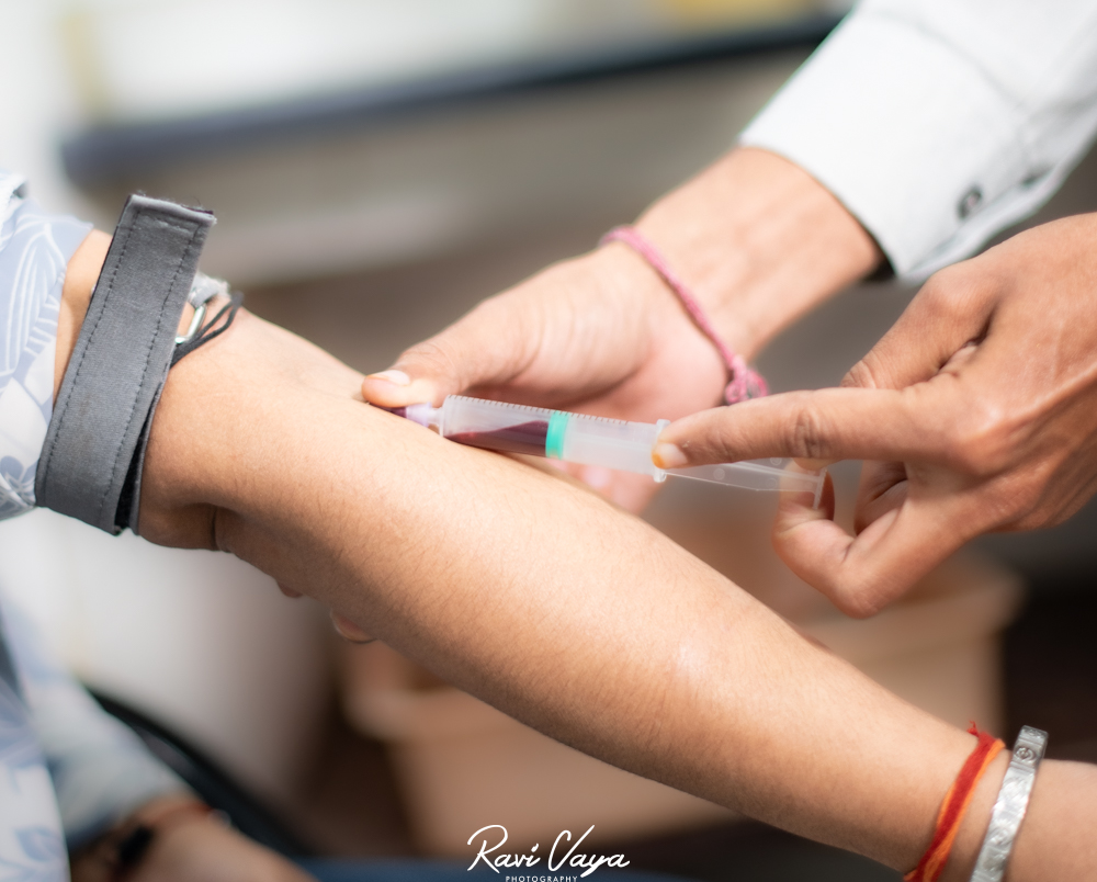
In the modern era of medical developments, diagnostic instruments are essential for precise medical assessment and early identification of possible health issues. The non-invasive imaging method known as ultrasound has grown to be essential to the diagnostics industry. But ultrasonography is more than just the blurry, conventional images you may be familiar with. Leading diagnostic centre in kota Dr. Vaya’s Lab provides an extensive array of cutting-edge sonography techniques that give unmatched detail and insights for a range of diagnostic needs.
This blog delves deeper into Doppler ultrasonography and 3D/4D sonography, two such advanced sonography techniques. We’ll go over their features, advantages, and the particular diagnostic applications they work well in, demonstrating Dr. Vaya’s Lab’s dedication to offering Kota residents state-of-the-art diagnostic services.
Doppler Ultrasound: Unveiling Blood Flow Dynamics
Conventional ultrasound imaging projects interior organs and structures onto a visual image by means of sound waves. This is further enhanced by Doppler ultrasound, which examines the direction and velocity of blood flow within these structures. This information is very helpful in the diagnosis of many circulatory system disorders.
How Does Doppler Ultrasound Work?
High-frequency sound waves are released by a specialized probe during a Doppler ultrasound examination. The Doppler effect causes these waves to change in frequency as they reflect off of red blood cells that are moving inside blood arteries. The ultrasound equipment then converts this change into colors or sounds, giving an aural or visual depiction of the direction and velocity of blood flow.
Benefits of Doppler Ultrasound:
- Early Detection of Cardiovascular Issues: Doppler ultrasound excels at identifying blockages or narrowing within arteries and veins (peripheral arterial disease), assessing blood flow to the heart muscle (coronary artery disease), and detecting abnormalities in heart valves.
- Evaluation of Blood Flow in Other Organs: Doppler ultrasound can be used to assess blood flow in the liver, kidneys, uterus, and other organs, aiding in the diagnosis of conditions like liver cirrhosis, kidney dysfunction, and abnormal uterine blood flow during pregnancy.
- Monitoring Fetal Well-being: Doppler ultrasound plays a crucial role in prenatal care by allowing doctors to evaluate blood flow to the fetus and placenta, helping to identify potential growth issues or complications.
3D/4D Sonography: Visualizing Anatomy in a New Dimension
Conventional ultrasound pictures display interior features in two dimensions. This restriction is overcome by 3D and 4D sonography techniques, which produce real-time visualizations and three-dimensional reconstructions, respectively.
- Three-dimensional sonography: This method makes use of several two-dimensional ultrasound pictures taken from different perspectives. These photos are subsequently processed by specialized software to produce an intricate 3D representation of the scanned organ or structure. With the help of this model, medical professionals can rotate, alter, and see the structure from various perspectives, giving them a more thorough grasp of its anatomy.
- 4D sonography: This technology builds on 3D technology by include time, allowing for the display of moving images in real time. This gives expectant parents a special bonding experience by enabling the visualization of fetal movement, breathing, and facial expressions in pregnant women.
Benefits of 3D/4D Sonography:
- Enhanced Diagnostic Capabilities: 3D sonography facilitates a more precise evaluation of complex anatomical structures, aiding in the detection of tumors, congenital abnormalities, and other anomalies that might be missed in a traditional 2D ultrasound.
- Improved Surgical Planning: 3D models generated from sonography can be invaluable for surgeons planning minimally invasive procedures, allowing for a more precise understanding of the surgical field.
- Early Detection of Fetal Abnormalities: 3D sonography can sometimes aid in the earlier detection of certain fetal abnormalities, allowing for prompt intervention if necessary.
- Enhanced Prenatal Experience: 4D sonography offers expecting parents a more realistic and emotionally connecting experience by allowing them to witness their baby’s movements in real-time.
Dr. Vaya’s Lab: Your Partner in Advanced Diagnostics
We are at Dr. Vaya’s Lab, a famous diagnostic centre in Kota. We dedicate ourselves to giving our patients access to the newest and best ways to diagnose. Our team uses modern Doppler ultrasonography and 3D/4D sonography equipment. Skilled radiologists and sonographers use this gear. It provides precise and educational results.
In order to meet your needs in a patient-centered manner, Dr. Vaya’s Lab is available for both standard exams and more involved evaluations. We make sure that the diagnostic process is quick and easy. And explaining the findings in detail and collaborating with your physician to determine the best course of action.